


in section Oncology
Tumors originating from the elements of the lymph node or extranodal lymphoid tissue are called lymphomas. The basis of modern histological and cytological classification of neoplastic diseases of the lymphoid and hematopoietic tissues laid the cellular composition of tumors. It is generally accepted international classification of cytologic and histologic tumor diseases of the hematopoietic and lymphoid tissue.
Malignant lymphoma - Lymphogranulomatosis (Hodgkin's disease)
 Among malignant lymphomasIt is about 5% of all cancers, accounting limfopranulematoza accounts 55-65%. Most often suffer from people aged 15-34 50 years and older.
The disease usually begins with the appearance of an enlarged, elastic, painless node in one of the groups of peripheral lymph nodes above the diaphragm. Gradually, other nodes of the same anatomical zone and then neighboring zones are involved in the process. The disease can be accompanied by common symptoms (weakness, night sweats, weight loss, fever, itching) or leak without them. The wave form of clinical manifestations is characteristic: the periods of exacerbation are replaced by periods of relative well-being. In the primary lesion of the mediastinal lymph nodes, the first clinical manifestation of the disease may be a syndrome of compression of the superior vena cava. Organic lymphogranulomatosis may be secondary, more rarely - primary. Most often, the lungs are affected secondarily, involvement in the process of the spleen and liver may not be accompanied by an increase in them, organ damage below the diaphragm is more often localized in the small intestine and stomach. With lymphogranulomatosis, bone and skin lesions become severe.
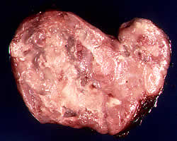
Hodgkin's disease diagnosis is based on morphological study of a pointed or remote enlarged lymph node. The presence of characteristic multinucleated cells Berezovsky-Sternberg natognomonichno for this disease.
The degree of spread of the process is determined by carrying out an X-ray examination of the chest organs, performing ultrasound of the abdominal cavity, excretory urography, and lymphography. The condition of the spleen is refined with the help of its radionuclide and angiographic studies. According to the indications, a radionuclide study of the bones of the skeleton is performed. The morphological investigation of the spleen, liver, lymph nodes and bone marrow occupies an important place in the staging of lymphogranulomatosis (see below). Therefore, the standard stage of examination of such patients is a diagnostic (explorative) laparotomy, during which splenectomy, biopsy of the liver and lymph nodes are performed. In young women, during this operation, the ovaries are sutured to the middle of the posterior surface of the uterus in order to remove them from the zone of subsequent intense irradiation. Trepanobiopsy also is mandatory in patients with lymphogranulomatosis.
Prognosis depends on the stage presence (category A) or absence (category B) symptoms of intoxication and the histological structure of the tumor. The most favorable option is to morphological lymphoid predominance of further deterioration in the forecast as follows nodular sclerosis, mixed-cell variant, lymphoid depletion.
The main method of treatment of patients with Hodgkin's disease is radiation therapy. For treatment uses sources of high energy radiation. The amount of exposure depends on the stage, histological types, as well as on the presence of systemic symptoms.
In the IA-IIA stages, irradiation of all lymphatic reservoirs is performed above the diaphragm, as well as the spleen and lumbar lymph nodes. 1B-NB stages carried out a total irradiation of the lymph nodes and spleen. In the ShA stage, irradiation of all lymph nodes, including nodes of the gates, liver and spleen is shown. In the stage of SB, irradiation of all organs of the abdominal cavity up to a dose of 20 Gy with protection of lead blocks of the kidneys after a dose of 12 Gy is possible; Then the irradiation continues as in the BOP stage. Usually, the irradiation is carried out with the figured fields first but one side of the diaphragm to the total dose of 40-44 Gy, then after the 4-6-week break, irradiation of the lymphatic collectors is performed on the other side of the diaphragm. In patients of Stage III, the irradiation can be performed by the method of an alternating split course, in which a dose of the order of 20-25 Gy is applied on one side of the diaphragm. Then, without interruption, also in a dose of 20-25 Gy the collectors are irradiated on the other side of the diaphragm, and then they return to irradiation of the first and then the second zone by 20 Gy for each. This technique allows only for 5 weeks to bring to all zones a dose of a radical level.
In patients with the presence of systemic symptoms of intoxication and / or organ damage is widely used subtotal body irradiation (CTOT) single focal dose 1,5 Gy up to a total dose of 4,5-6,0 Gy. Such exposure is an alternative to systemic chemotherapy.
Radiation therapy is the only treatment only at stage I-IIA in the case of prognostically favorable morphological variants. Sometimes during these stages used chemoradiotherapy.
Chemotherapy is also directed to methods of treatment of patients with primary Hodgkin's disease. Monochemotherapy rarely used, mainly in elderly patients, as well as during the process of continuously recurrent. Chemotherapy is widely used in the treatment of patients with primary, in generalized form, as well as the original local process in combination with irradiation. In terms of self-chemotherapy is indicated for 4V stage. A distinctive feature of antitumour therapy in the treatment of Hodgkin -mnogokursovoe.
The most effective treatments are considered TSOPP (cyclophosphamide, Oncovin, procarbazine, prednisone) OPP (the same without cyclophosphamide); MOTAPMs (mustargen, Oncovin, procarbazine, prednisone) WFP (mustargen, procarbazine, prednisone) AIC ((mustargen, adriamycin, bleomycin, Oncovin) TSVPP (cyclophosphamide, vinblastine, natulan prednisolone) ABDV (adriamycin, bleomycin, dacarbazine, vinblastine), and others.
Chemoradiotherapy - the main type of treatment of Hodgkin's disease. At stage II-IIIA treatment is usually started with 3-6 courses of chemotherapy, the second stage is used for radiation therapy regimens described above, is then carried out for at least three courses of chemotherapy.
Currently, indicators 5-year survival in patients with Hodgkin's disease, depending on the stage range from up to 40-55 80-90%. Patients who have lived 5 years without recurrence have 95% chance of definitive cure.
Malignant Lymphoma - Lymphoma
Lymphoma - a malignant tumor, morphological substrate of which are elements of the lymphoid cell, is clinically characterized by lymph nodes and various organs, and hematogenous metastasis lymphogenous with leukemization in 20% of patients. In the structure of malignant hematological malignancies at a fraction lymphosarcoma accounts for about 15%, the frequency they are second only to Hodgkin's disease. In most cases, lymphosarcoma has B-cell origin, T-cell variants are much less common.
The main histological forms of lymphosarcoma are nodular; lymphocytic, lymphoplasmacytic, prolymphocytic, immunoblastic and lymphoblastic.
The clinic depends on the location of the tumor, in most cases the disease begins with an increase in the peripheral lymph node or group of nodes. With organ lesions, the symptoms correspond to the tumor lesion of this organ. The diagnosis of lymphosarcoma is mainly histological. With the morphological confirmation of the diagnosis, an x-ray examination of the organs of the chest, gastrointestinal tract, radionuclide evaluation of bones, liver and spleen, ultrasound and computer evaluation is performed. Mandatory is trepanobiopsy of the bone marrow and the study of the myelogram. Laparotomy and splenectomy with lymphosarcoma, given the frequent hematogenous metastasis, are not performed.
For lymphosarcoma characterized by rapid tumor involvement in the process of extranodal organs and tissues, particularly the bone marrow that affects at 20% of patients with the development of leukemia. Lymphosarcoma often metastasize to the liver (20-40%), second lien involved in the process from the 20-30% of patients. Primary tumor localization observed in the elbow, genial, occipital areas, which is not seen with chlamydia.
The main method of treating lymphosarcoma is chemo-therapeutic. Most antitumor drugs alkylating, antimetabolites, antibiotics, herbal preparations, corticosteroids - are used to treat lymphosarcomas. Monochemotherapy (cyclophosphamide, chlorbutin, vincristine) causes remission in 30-60% of patients, with complete regression of tumor foci only achieved in 10-20% of observations. The oncoming remissions are short-lived - 1-4 months. The use of two or more cytostatics allows direct effect in 75-95% of patients with lymphosarcomas, the duration of remission increases to 15-45 months. The most common schemes of polychemotherapy with lymphosarcoma are cyclophosphamide, vincristine, prednisolone; Vincristine, methotrexate, 6-mercaptopurine, prednisolone; Cyclophosphamide, vincristine, prednisolone, bleomy, etc.
Radiation therapy is used primarily in the I-II stage of the process and allows you to achieve complete tumor resorption in 40-60% of patients. The best results are achieved by radiotherapy lymphosarcoma in the defeat of the tonsils and throat. Methods of exposure in local populations is to irradiate the affected lymph nodes and regional areas. In normal mode, the total focal dose fractionation are brought to 40-45 Gy.
Surgery for lymphosarcoma is of limited value, but the localization of tumors in the gastrointestinal tract, testes, thyroid and breast cancer surgery not only possible, but also leads to prolonged healing 30-40% of patients.
The most effective for today is the chemoradiation treatment of patients with lymphosarcomas. As a rule, at the initial stages of the disease, the first stage of the irradiation of the affected areas, followed by a course of polychemotherapy, listed above. In cases of a common disease, treatment starts with chemotherapy, and then residual tumor lesions are irradiated. Combined chemoradiotherapy allows to achieve complete regression of tumors in 85-88% of patients, the average duration of remission in this case exceeds 40 months. At 1-P stage, the five-year survival of patients after chemoradiotherapy in some clinics reaches 80-85%.
Malignant lymphoma - Retnkulosarkoma
Retikulosarkome (gistioblastnaya malignant lymphoma) - a malignant tumor that is the morphological substrate gistiotsit. Clinic and diagnostics retikulosarkom no different from that in lymphosarcoma. Retikulosarkome occur at any age, is more common in men. In the early stages of the disease is marked local lymph nodes, spleen, bones and other organs. With the progression of the process is marked infiltrative growth. Generalization occurs by metastasis to distant organs and tissues.
Clinical manifestations of the disease are varied and depend on the location of the tumor and the extent of its spread. Retikulosarkome infiltrates the underlying tissue, skin, blood vessels and nerves grows and causes unbearable pain. With involvement of the bone marrow is its leukemization with the development of acute myeloid paintings, rarely gistiomonoblastnogo leukemia.
The main method of diagnosis is histological examination of the tumor. The discovery of large reticular cells or atypical polymorphic reticular cells favors rstikulosarkomy. Diagnosis of the extent of the process carried out by the same methods as for lymphosarcoma.
The main treatment is chemotherapy reticulosarcoma. Apply the same drugs and schemes that for the treatment of lymphosarcoma, but preferred alkylating agents (sarkolizin, cyclophosphamide) and a combination of anthracyclines (rubomycin, adriamitsii) with cytosine arabinoside. Application nolihimioterapii leads to the achievement of the objective effect in 50- 70% of patients, but the duration of remission is low and is only a few months.
Clasmocytoma sensitive to radiation therapy, which is used as a component of an integrated treatment, generally at a 1-II stage. Total focal doses are brought to 50-60 Gy.
Surgical treatment retikulosarkom usually conducted at the tumor site in the gastrointestinal tract.
Forecast at retikulosarkome unfavorable, the average life expectancy of patients with no more than two or three years.
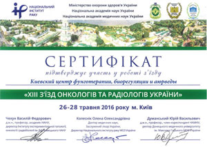
В Kiev Center for Fungotherapy, Bioregulation and Ayurveda qualified doctors of alternative medicine are receiving. Cost of consultation 300 UAH. You can look at medical histories and treatment results on this link.
You can make an appointment by phone: (097) 231-74-44, (050) 331-74-44, (063) 187-78-78, +38 (098) 583-85-85 (Viber), +38 (093) 688-25- 88 (WhatsApp, Telegram) Email:This email address is being protected from spambots. You need JavaScript enabled to view it.


 Many messenger users use the Telegram application, as, unlike Viber, new members of the group can see the previous correspondence in the group. To join the "Alternative Medicine" group in Telegram, scan the QR code or follow the invitation link


")

")