


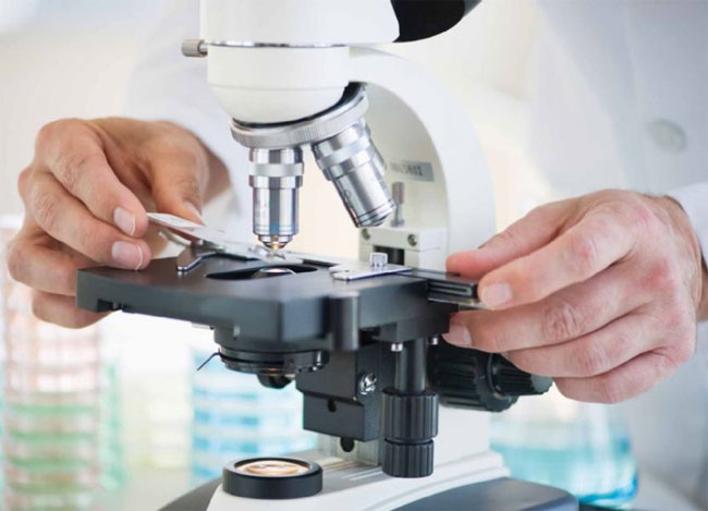
The method of microscopy of native blood is a new and interesting study of the inner environment of a person, unique in its accessibility, ease of carrying out, informative and important for the patient.
Blood is an amazing creation of nature. It can be no exaggeration to say that it is the source of life.
Blood constantly circulates through our body and without this movement life is simply impossible. It penetrates into all organs and tissues and can change the composition depending on the state of the organism. That's why one blood test can often get information about the transferred and existing diseases, the general condition of the body and violations in different organs.
Blood was one of the first liquids that curious naturalists put under the newly invented microscope. Since then, more than 300 years have passed, and interest in studying it has not disappeared to this day. There are many methods that allow you to obtain data on different states of the blood and to judge them about changes in the body.
In laboratory practice, microscopy of a stained smear is most often used. To do this, the blood smear is pre-dried, fixed and colored, and then after a certain time, the cells are counted, their structure and appearance are described. Native or "living" blood (without fixation and coloration) is microscopically rare. But the study of a "live" drop is the simplest and most informative method of research, which has been known for a long time and has been widely used in the past centuries. Due to different circumstances, medical practice has departed from the daily use of this method, but today in the era of computers and digital technologies attention to the method of research of native blood is again increasing and is gaining popularity.
The microscope could be connected to a digital video camera and a computer, which made it possible to significantly increase Visualize blood objects on the screen, difficult to distinguish in a conventional light microscope, with the possibility of saving the image for further work. Such a hardware complex allows not only to view the cellular elements of the blood, but also to evaluate their dynamic functional characteristics, the purity of the plasma and the presence of parasitic forms (bacteria, fungi, protozoa without their species identification), and also demonstrate testing to the patient. In this regard, the patient takes a direct part in the process, has a unique opportunity to see his cells during the study, obtains important information for him and has the opportunity to assess his own condition, lifestyle, to participate in cooperation with the doctor in matters of restoring his health.
Blood is considered the river of life. And like any river, it has its own population. Each component of this river has its own functions, thanks to which our organism exists. This transport, nutritional, protective functions, providing oxygen and organs and tissues, maintaining the pH level of blood and body fluids and much more. Between the blood cells and the cells of the organs, information and energy interchange constantly occurs, which is possible with a certain qualitative level of functioning of enzyme systems, and in turn, they need macro-, microelements, vitamins, oxygen, mitochondrial energy and a qualitative liquid medium. In a living drop of blood, we are able to observe the interaction of plasma and cells, the state of cells and processes occurring in the plasma. As a result, sludge syndrome (adhesion of erythrocytes into cellular conglomerates), anemia syndromes, intoxication, dysfunction of leukocytes, risk of increased thrombus formation, indirectly judge the water deficit in tissues, the presence of inflammation, dysbacteriosis, etc. can be detected.
Therefore, the study of a live drop of blood, which is a kind of "mirror" of the body, can give extremely important information about the health status in real time, as well as possible problems that may arise as a result of prolonged disruption of homeostasis. Thus, the condition of the pre-illness can be detected long before the development of the disease.
The method of microscopy of native blood has its own specific capabilities and specific goals that correspond to the following:
- determination of non-quantitative levels of saturation with oxygen, water and necessary nutrients for the body;
- the study of the ratio of plasma and cellular elements, the degree of adhesion of red blood cells, which is reflected in the fluidity of the blood;
- Assessment of the coagulation system (propensity to thrombosis and prognosis of complications associated with it);
- analysis of the morphological characteristics of erythrocytes (shape, size, intensity of color), which can be used to judge the presence of anemia and somatic diseases;
- causes of damage to red blood cells - lack of folic acid and vitamin B12, iron deficiency;
- degree of purity of blood plasma and various inclusions: cholesterol, sugar, urate salts;
- Evaluation of the state of leukocytes, but according to their functional activity of the entire immune system;
- a study of the purity of plasma for the presence of biological objects - bacteria, fungi, other microorganisms without their identification;
- definition of signs of inflammatory processes, including hidden ones;
- Definition of markers of cardiovascular diseases;
- the stage of accumulation and distribution of slags in the human body;
- evaluation of the body's functional reserves;
- the state of fat metabolism and predisposition to atherosclerotic processes.
Thus, the study of native blood is a serious tool in testing the functional state of the body, identifying predispositions to specific diseases, creating recovery and health programs.
Typically, the study of blood is done in two stages. (Before carrying out the analysis, it is advisable to refrain from eating for 2-3 hours). At the first stage, a primary examination under a microscope of extracted blood from the finger (just one drop is enough). After this, if there are indications, the patient is offered to drink 200 - 300 ml of warm water with the addition of the necessary preparation for the study and a second study is carried out.
In our Center of Fungotherapy, biregulation and ayurveda, the method of native blood microscopy can be used both to check the general condition of the body and early diagnosis of diseases, and to visualize the effect on the blood state, and therefore on the entire body, of certain medications.
That is, the patient on the monitor screen can observe the blood picture before and after taking the drug, and get professional advice from a doctor with comments on this process. Thus, it can be determined whether the patient is suitable for this drug or not, see the activation of the immune system, the effect of the drug on the degree of adhesion of erythrocytes and other processes.
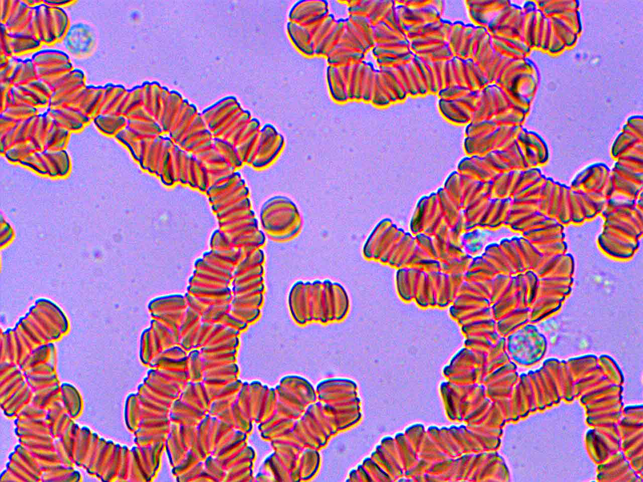

The degree of adhesion of red blood cells before and after taking Rain Soul
The method of microscopy of native blood greatly helps to assess the general condition of the patient's body, the propensity to develop certain diseases and, as a consequence, correctly determine the effective treatment regimen with the following recommendations for nutrition and the use of natural biological supplements and medicines.
The cost of diagnostics is 150 UAH
В Kiev Center for Fungotherapy, Bioregulation and Ayurveda qualified doctors of alternative medicine are receiving. Cost of consultation 300 UAH. You can look at medical histories and treatment results on this link.
You can make an appointment by phone: (097) 231-74-44, (050) 331-74-44, (063) 187-78-78, +38 (098) 583-85-85 (Viber), +38 (093) 688-25- 88 (WhatsApp, Telegram) Email:This email address is being protected from spambots. You need JavaScript enabled to view it.

 Many messenger users use the Telegram application, as, unlike Viber, new members of the group can see the previous correspondence in the group. To join the "Alternative Medicine" group in Telegram, scan the QR code or follow the invitation link


")